

In 1993 DICOM has revolutionized the healthcare industry. What was once a chaotic landscape riddled with multiple proprietary image formats and communication protocols has transformed into a standardized, interoperable ecosystem. Thus, we couldn’t overlook DICOM in our series of interviews about the most important standards across industries. So, let’s uncover what DICOM is and how exactly it drives healthcare forward.
What is DICOM?
DICOM, short for Digital Imaging and Communications in Medicine, is a global standard for medical images and the associated information. It sets the formats for medical images that can be shared, as well as the quality and data requirements necessary for clinical applications.
DICOM is employed in nearly all radiology, cardiology, and radiotherapy equipment, including ultrasound, CT scans, MRI, and others. It’s also used in various medical devices in other fields like dentistry and ophthalmology. Published by the National Electrical Manufacturers Association (NEMA), DICOM stands out as one of the most widely utilized standards for message exchange in the global healthcare industry.
Since its introduction, DICOM has transformed radiology practices, completely replacing X-ray film with a digital workflow. As the internet evolved into a platform for new consumer information applications, DICOM made cutting-edge medical imaging applications accessible, fundamentally altering the landscape of clinical medicine. From emergency room care to stress testing and cancer cell detection, DICOM serves as the standard that enables medical imaging to benefit both healthcare professionals and patients.
DICOM is recognized by the International Organization for Standardization as ISO 12052 (with the latest revision in 2017). The standard is regularly updated, sometimes with multiple updates in a single year. For instance, in 2021 the standard saw two updates, and in 2020, there were five updates.
What is the structure of DICOM?
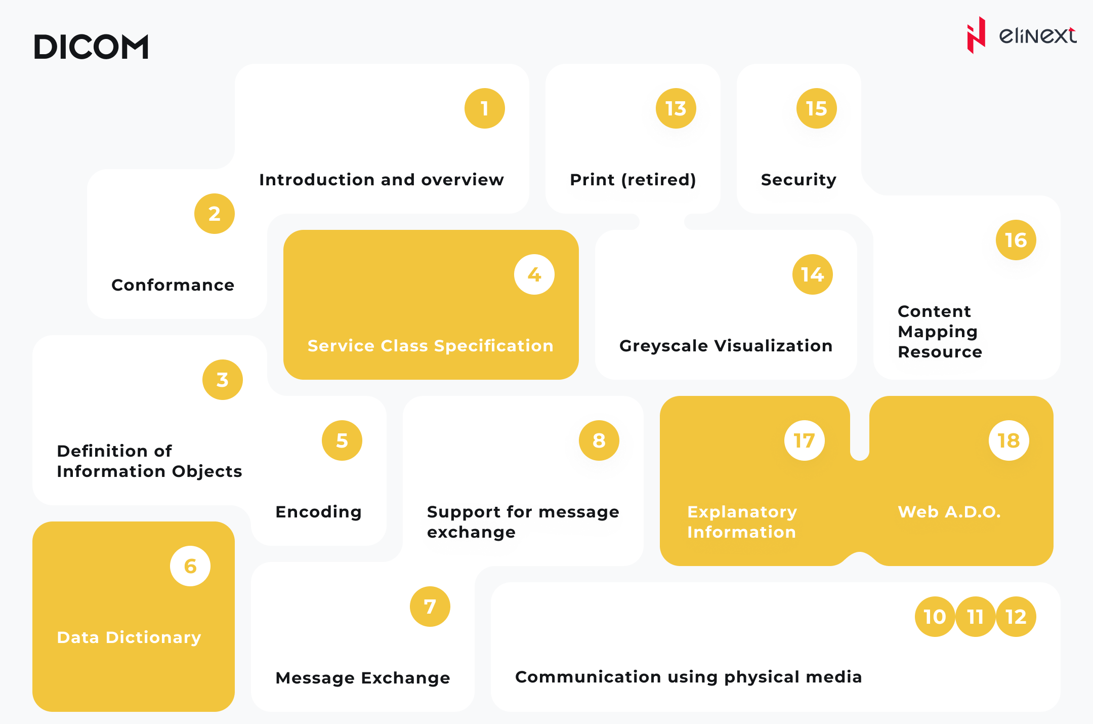
DICOM doesn’t just define sets of images and data; it also covers the transport protocol and various other services. As of now, the entire standard comprises 22 interconnected yet standalone sections. For instance, there are sections that encompass:
- handling procedure worklists for visualization purposes;
- transferring images to digital media, such as DVDs;
- generating reports on procedure status, including the completion of image acquisition;
- verifying the successful image archiving;
- securing data sets through encryption;
- anonymizing patient-identifying information;
- structuring image layouts for viewing;
- arranging image layouts for viewing;
- preserving image manipulations, annotations, and more.
What is the purpose of DICOM?
Well, DICOM is practically a universal standard in hospitals around the world. It ensures the compatibility of systems used for creating, storing, sending, displaying, and otherwise processing of medical images and structured documents. DICOM also helps manage the associated workflow.
Who benefits from DICOM?
DICOM’s primary objective is to meet the requirements of the following stakeholders:
- Healthcare professionals
DICOM provides doctors with speedy access to images and reports, enabling quicker diagnoses.
- Patients
Patients receive faster and more efficient care when medical facilities use DICOM to share information.
- Hospitals, clinics, imaging centers, and their specialists
This group can make use of DICOM compliance as part of their purchasing criteria, ensuring seamless compatibility between the acquired equipment and software solutions.
- Manufacturers of image processing equipment, image processing information systems, and peripheral equipment.
What are DICOM’s key concepts?
To better understand DICOM standard, let’s have a look at the main concepts.
Data structure
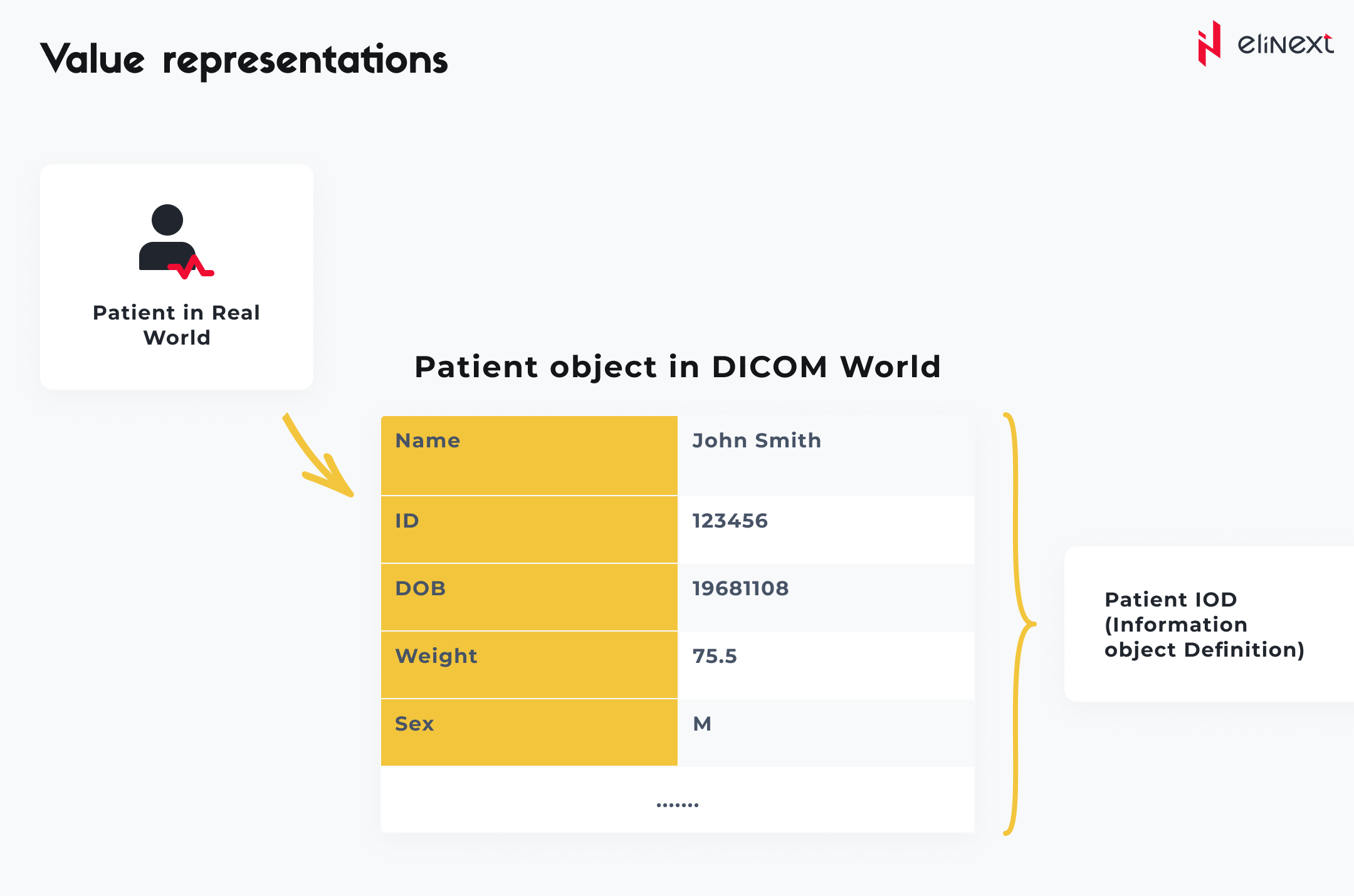
DICOM arranges data into datasets. This means that a file containing, for instance, a chest X-ray, actually includes the patient’s identifier within the file, preventing any accidental detachment of this crucial information from the image. This concept is akin to image formats like JPEG, which can also embed tags for image identification and other descriptions.
Image display
In order to achieve consistent grayscale image rendering on various monitors and uniform printed image quality across different printers, the DICOM committee has created a reference table for mapping digitally assigned pixel values. To utilize the standard grayscale display function of DICOM images, it’s essential to view and print them on devices equipped with this lookup curve or on devices calibrated to adhere to the grayscale curve.
Value representations
Beyond simply representing the value, each attribute also defines a value multiplicity to signify the number of data elements contained within the attribute. When dealing with string and character values, in cases where multiple data elements are encoded, they are separated by a backslash symbol.
Services
DICOM encompasses various services, with most of them revolving around network data transmission. The ile format for offline media was added later to the standard.
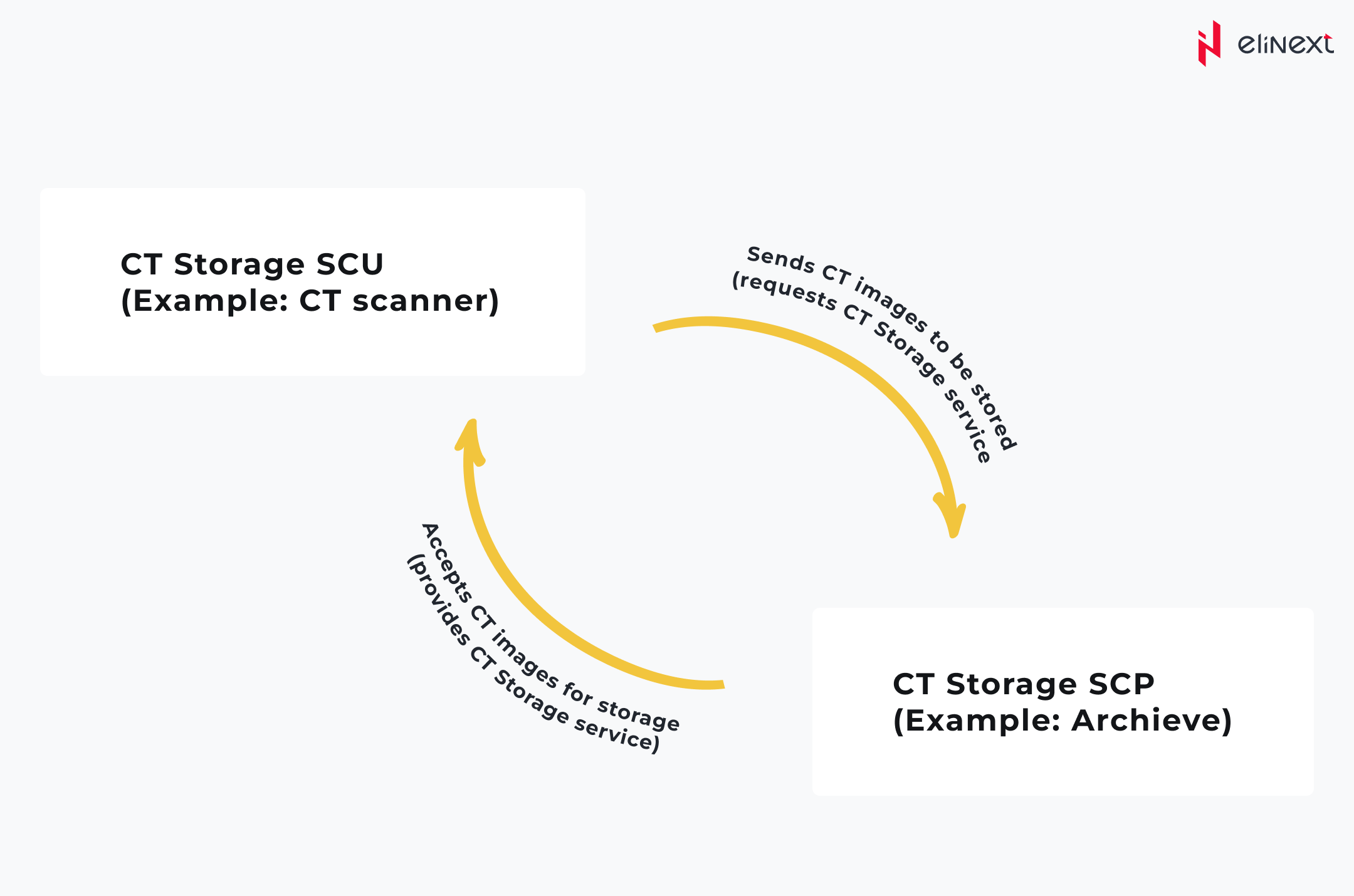
Storage
The DICOM storage service facilitates the transmission of images or other permanent objects to a Picture Archiving and Communication System (PACS) or a workstation.
Storage Commitment
The DICOM Storage Commitment service is employed to validate the storage of images, either on the device or on backup disks or media (e.g., as recorded on a compact disc).
Queries
Queries empower a workstation to search for lists of images or similar objects within the PACS and subsequently retrieve them.
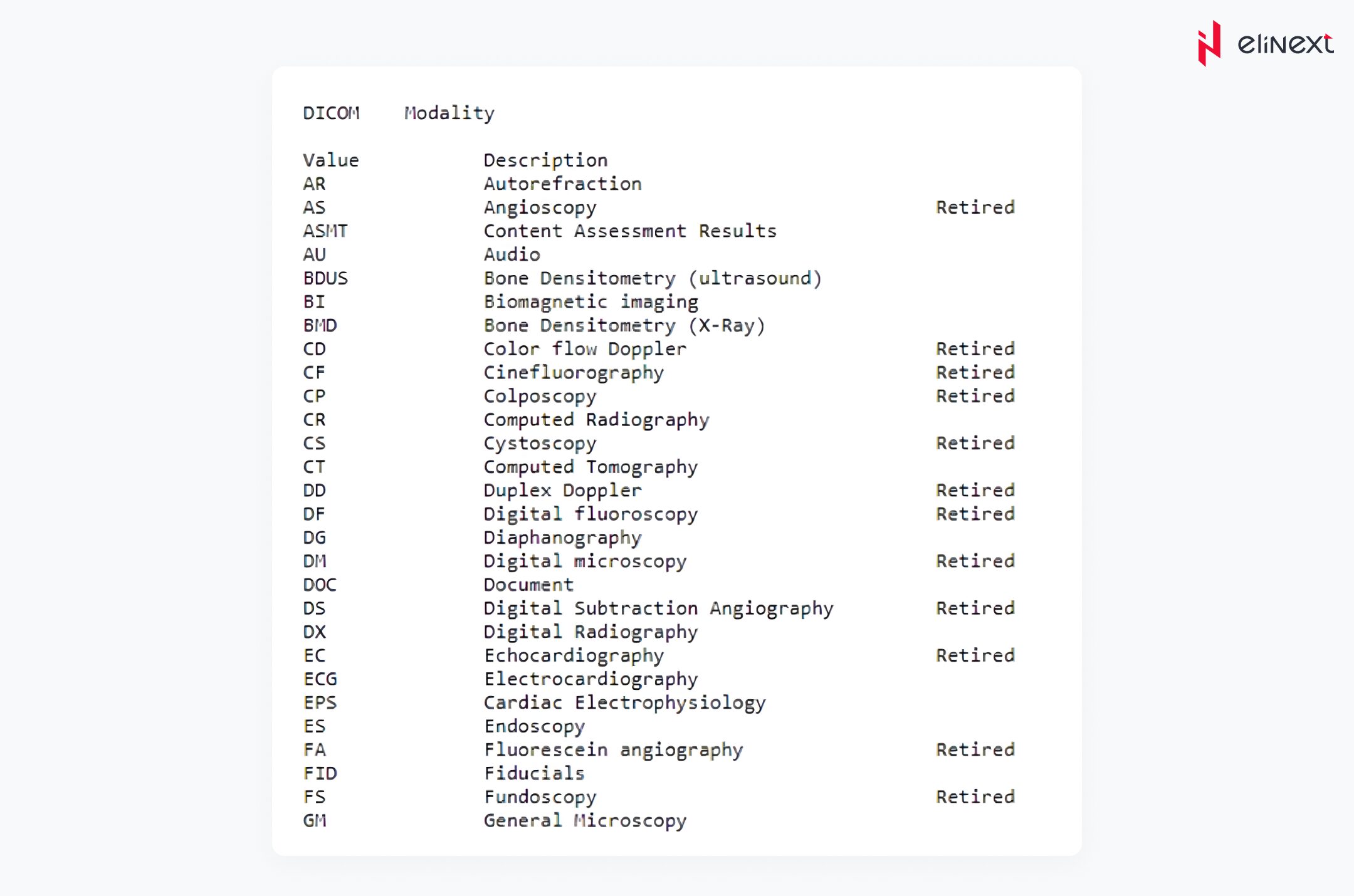
What is a modality in the context of DICOM?
A DICOM data object comprises a set of attributes, including elements such as the patient’s name and identifier, as well as attributes containing information about pixel images. One of these attributes is the modality, which represents the file type. You can also think of it as the type of equipment used to obtain the file. DICOM establishes a functional list of modalities.
Modality Worklist
This service offers a list of image processing procedures scheduled for execution by the image acquisition device. The worklist elements encompass corresponding details about the procedure subject, type, order, and diagnostic reason.
Modality Performed Procedure Step (MPPS)
MPPS enables modalities to submit a report about the performed examination, encompassing details on acquired images, start and end times, examination duration, administered dosage, and more. MPPS also helps modalities in enhancing the coordination of their actions with image storage servers. It provides the server with a list of objects to transmit before or during the actual transmission of these objects, allowing for more precise resource management in departments like radiology.
Printing
This service is employed for dispatching images to a DICOM printer, usually for the purpose of printing X-ray films. A standardized calibration ensures uniformity across diverse display devices.
Offline media
The format for standalone multimedia files is outlined in Part 10 of the DICOM standard. DICOM limits file names on DICOM media to 8 characters, providing no informational clues from these names. DICOM files usually sport the .dcm extension, unless they are part of a DICOM media, in which case they go without an extension.
Just like any standard, DICOM has its own benefits and limitations. What are they?
Indeed, DICOM offers multiple benefits to medical services, including:
● Collaboration with existing IT systems
In some cases, the incompatibility and improper image storage methods for the pathologies we are dealing with can be a significant hurdle when transitioning to digital pathology. The introduction of digital pathology took place as early as 2010 and was well-received by hospitals because it allowed them to integrate digital pathology into their existing systems without incurring any extra costs. The standard allows for the storage of pathology images along with other images in a unified archive.
● Highly efficient analysis
The extensions in which DICOM images are stored demonstrate consistently high performance every time the images are viewed. The scanner’s highest resolution captures the most data.
● Comprehensive image scanning and viewing
The majority of scanners don’t capture the entire slide at the highest resolution. These images, often referred to as sparse, can be effectively handled using the DICOM standard. This enables the removal of uncaptured areas from the images and reduces the image size, resulting in faster scanning times.
● Accurate image and patient information storage
With DICOM, patient information within an image is stored using tags. Any system reading these images can access and display the data seamlessly.
As for DICOM’s limitations, the most notable are:
● Slow scanning
Scanners are usually fine-tuned for speed, but if they haven’t been updated to meet the DICOM standard, additional steps during scanning may be necessary, potentially extending the scanning time.
● Indexing
To ensure efficient viewing, indexing is necessary
● DICOM header
The DICOM header consists of multiple tags, some of which are optional, while others are required. Scanner manufacturers may face difficulties in selecting these tags, potentially leading to the risk of unreadable information, such as patient data or other essential details getting lost.
Is DICOM certification mandatory and how can one obtain it?
DICOM certification is not obligatory and NEMA lacks the authority and obligation to oversee or guarantee compliance with the DICOM standard. In NEMA’s disclaimer, it’s explicitly mentioned that product sellers, including hardware or software developers, essentially confirm that their products align with the DICOM standard when they use the DICOM label for their products. In simple terms, manufacturers are claiming self-certification or compliance with these standards.
However, there is one organization called PARCA (PACS Administrators Registry and Certification) that provides DICOM certification for professionals since 2005. Certified DICOM Integration Professional, PARCA’s certification, is aimed at a diverse audience, including PACS system administrators, integration specialists, procurement and specification agents, technical imaging experts, informatics professionals, service engineers, software developers, and advanced system administrators/managers.
A certification from PARCA, Certified DICOM Integration Professional, is designed to cater to a broad spectrum of professionals. This includes PACS system administrators, procurement and specification agents, technical imaging experts, service engineers, software developers, system administrators, managers, and others.
The purpose of this certification is to assess and support the ability to connect DICOM, troubleshoot issues, carry out integrations, and build software solutions. The certification remains valid for 5 years, and for re-certification, participants need to maintain an active membership and re-take the exam.
Wrapping up
In the realm of healthcare, DICOM emerges as a crucial pillar that supports compatibility and seamless integration among a wide range of medical imaging devices and systems. As such, DICOM has found acceptance among healthcare providers, laboratories, and medical facilities, allowing for efficient sharing of diagnostic information and contributing to the advancement of patient care.
If you are looking for a trusted software development partner for your next healthcare project, we got you covered. At Elinext, we have accumulated significant expertise in developing interoperable custom healthcare solutions that meet the strictest industry standards, including DICOM, HL7 FHIR, HIPAA, PIPEDA, PHIPA, and others.
